Advanced Analytical Centre Analytical Facilities All Instruments Electron Probe Microanalyser (EPMA or Microprobe)
Electron Probe Microanalyser (EPMA or Microprobe)
- Future Students
- JCU Global Experience
- International Students
- Open Day
- How to apply
- Pathways to university
- Virtual Open Day
- Living on Campus
- Courses
- Publications
- Scholarships
- Parents and Partners
- JCU Heroes Programs
- Aboriginal and Torres Strait Islander in Marine Science
- Elite Athletes
- Defence
- Current Students
- New students
- JCU Orientation
- LearnJCU
- Placements
- CEE
- Unicare Centre and Unicampus Kids
- Graduation
- Off-Campus Students
- JCU Job Ready
- Safety and Wellbeing
- JCU Prizes
- Professional Experience Placement
- Employability Edge
- Art of Academic Writing
- Art of Academic Editing
- Careers and Employability
- Student Equity and Wellbeing
- Career Ready Plan
- Careers at JCU
- Partners and Community
- JCU-CSIRO Partnership
- Alumni
- About JCU
- Reputation and Experience
- Chancellery
- Governance
- Celebrating 50 Years
- Academy
- Indigenous Engagement
- Education Division
- Graduate Research School
- Research and Teaching
- Research Division
- Research and Innovation Services
- CASE
- College of Business, Law and Governance
- College of Healthcare Sciences
- College of Medicine and Dentistry
- College of Science and Engineering
- CPHMVS
- Anthropological Laboratory for Tropical Audiovisual Research (ALTAR)
- Anton Breinl Research Centre
- Agriculture Technology and Adoption Centre (AgTAC)
-
Advanced Analytical Centre
- About us
- Commercial and external clients
-
Analytical Facilities
-
All Instruments
- X-ray Powder Diffraction (XRD)
- X-ray Fluorescence (XRF)
- Scanning Electron Microscopy (SEM)
- Inductively Coupled Plasma-Mass Spectrometer (ICP-MS)
- Laser ablation
- Inductively Coupled Plasma-Atomic Emission Spectrometer (ICP-AES)
- Gas Chromatography-Liquid Chromatography (GC/LC)
- Additional Equipment
- Laser Scanning Confocal Microscopy (LSCM)
- Advanced Analytical Centre
- Multicollector-Inductively Coupled Plasma-Mass Spectrometer (MC-ICP-MS)
- Electron Probe Microanalyser (EPMA or Microprobe)
- Sample Requirements
- Techniques and Facilities
-
All Instruments
- Staff
- Safety
-
Resources
- Element-to-stoichiometric oxide conversion factors
- Gunshot residue (GSR)
- FAQ ICP
- FAQ Organic
- SEM images of local insects – N.Queensland
- False coloured images
- Notes on sample preparation of biological material for SEM
- Routine XRF element analysis @ AAC
- How to view/edit element maps from Jeol 8200 EPMA
- Standard ICP element analysis @ AAC
- FAQ - XRD/XRF
- Other JCU Facilities
- Contact the AAC
- AMHHEC
- Aquaculture Solutions
- AusAsian Mental Health Research Group
- ARCSTA
- Area 61
- Lions Marine Research Trust
- Australian Tropical Herbarium
- Australian Quantum & Classical Transport Physics Group
- Boating and Diving
- Clinical Psychedelic Research Lab
- Centre for Tropical Biosecurity
- Centre for Tropical Bioinformatics and Molecular Biology
- CITBA
- CMT
- Centre for Disaster Solutions
- CSTFA
- Cyclone Testing Station
- The Centre for Disaster Studies
- Daintree Rainforest Observatory
- Fletcherview
- JCU Eduquarium
- JCU Turtle Health Research
- Language and Culture Research Centre
- MARF
- Orpheus
- TESS
- JCU Ideas Lab
- TARL
- eResearch
- Indigenous Education and Research Centre
- Estate
- Work Health and Safety
- Staff
- Discover Nature at JCU
- Cyber Security Hub
- Association of Australian University Secretaries
- Services and Resources Division
- Environmental Research Complex [ERC]
- Foundation for Australian Literary Studies
- Gender Equity Action and Research
- General Practice and Rural Medicine
- Give to JCU
- Indigenous Legal Needs Project
- Inherent Requirements
- IsoTropics Geochemistry Lab
- IT Services
- JCU Webinars
- JCU Events
- JCU Motorsports
- JCU Sport
- Library
- Mabo Decision: 30 years on
- Marine Geophysics Laboratory
- Office of the Vice Chancellor and President
- Outstanding Alumni
- Pharmacy Full Scope
- Planning for your future
- Policy
- PAHL
- Queensland Research Centre for Peripheral Vascular Disease
- Rapid Assessment Unit
- RDIM
- Researcher Development Portal
- Roderick Centre for Australian Literature and Creative Writing
- Contextual Science for Tropical Coastal Ecosystems
- State of the Tropics
- Strategic Procurement
- Student profiles
- SWIRLnet
- TREAD
- TropEco for Staff and Students
- TQ Maths Hub
- TUDLab
- UAV
- VAVS Home
- WHOCC for Vector-borne & NTDs
- Media
- Copyright and Terms of Use
- Australian Institute of Tropical Health & Medicine
- Pay review
Technique in brief
An Electron Probe Microanalyser (EPMA) combines the features of a scanning electron microscope (high magnification, high resolution imaging) with elemental analysis at the micron scale. It allows for the non-destructive analysis of very small amounts of solid materials. As with a SEM, an electron beam is scanned across the surface of a sample and the interaction of the beam electrons with the atoms of the material examined produce a number of measurable effects. As well as producing imaging information X-rays are generated from the sample. By measuring the energy or wavelength of these X-rays the elements present can be identified. Comparison of the intensity of X-rays produced against that of standards of known composition allows for this data to be quantified.
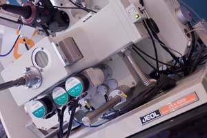
Current instrumentation
The AAC currently houses a Jeol JXA8200 “Superprobe” equipped with:
5 wavelength dispersive spectrometers (WDS), each with dual crystals
Energy dispersive spectrometer (EDS)
Tungsten (W) and LaB6 electron gun
Backscatter electron imaging (BEI)
Secondary electron imaging (SEI)
Cathodoluminescence (CL) - wavelength spectrometer system (XCLent)
High speed, high resolution, large area stage
Applications
An EPMA can be used in many areas of research, principally when chemical analyses are required on the micron scale (ie individual grains or small areas rather than bulk/whole sample chemistry). It is capable of elemental analysis from B to U with typical analysis volumes between 1 and 3 microns. In addition to single spot analysis, line traverses (eg for core to rim analysis of a mineral) or element maps can be produced. In the latter a grid of analysis are run and the spatial distribution of element distribution can be displayed as an image (map).
Some sample types produce visible light when excited by an electron beam; this is known as cathodoluminescence (CL). Variation in CL is often the result of changes in trace element chemistry or structural deformation/imperfections and can thus be used to distinguish between different generations of a mineral phase. The EPMA at the AAC is equipped with an XCLent wavelength spectrometer based CL system. Used in conjunction with element mapping, this can produce images illustrating the spatial distribution of CL intensity along with the variations in actual wavelengths of light being produced.
Sample requirements
As for SEM, samples for EPMA need be stable under vacuum and, if not electrically conductive, coated (typically for analysis carbon is used). For quantitative analysis they need to have a flat, polished surface (typically a polished thin section or resin mount).
For further information contact one of the following officers in charge: