Advanced Analytical Centre Analytical Facilities All Instruments Laser Scanning Confocal Microscopy (LSCM)
Laser Scanning Confocal Microscopy (LSCM)
- Future Students
- JCU Global Experience
- International Students
- Open Day
- How to apply
- Pathways to university
- Virtual Open Day
- Living on Campus
- Courses
- Publications
- Scholarships
- Parents and Partners
- JCU Heroes Programs
- Aboriginal and Torres Strait Islander in Marine Science
- Elite Athletes
- Defence
- Current Students
- New students
- JCU Orientation
- LearnJCU
- Placements
- CEE
- Unicare Centre and Unicampus Kids
- Graduation
- Off-Campus Students
- JCU Job Ready
- Safety and Wellbeing
- JCU Prizes
- Professional Experience Placement
- Employability Edge
- Art of Academic Writing
- Art of Academic Editing
- Careers and Employability
- Student Equity and Wellbeing
- Career Ready Plan
- Careers at JCU
- Partners and Community
- JCU-CSIRO Partnership
- Alumni
- About JCU
- Reputation and Experience
- Chancellery
- Governance
- Celebrating 50 Years
- Academy
- Indigenous Engagement
- Education Division
- Graduate Research School
- Research and Teaching
- Research Division
- Research and Innovation Services
- CASE
- College of Business, Law and Governance
- College of Healthcare Sciences
- College of Medicine and Dentistry
- College of Science and Engineering
- CPHMVS
- Anthropological Laboratory for Tropical Audiovisual Research (ALTAR)
- Anton Breinl Research Centre
- Agriculture Technology and Adoption Centre (AgTAC)
-
Advanced Analytical Centre
- About us
- Commercial and external clients
-
Analytical Facilities
-
All Instruments
- X-ray Powder Diffraction (XRD)
- X-ray Fluorescence (XRF)
- Scanning Electron Microscopy (SEM)
- Inductively Coupled Plasma-Mass Spectrometer (ICP-MS)
- Laser ablation
- Inductively Coupled Plasma-Atomic Emission Spectrometer (ICP-AES)
- Gas Chromatography-Liquid Chromatography (GC/LC)
- Additional Equipment
- Laser Scanning Confocal Microscopy (LSCM)
- Advanced Analytical Centre
- Multicollector-Inductively Coupled Plasma-Mass Spectrometer (MC-ICP-MS)
- Electron Probe Microanalyser (EPMA or Microprobe)
- Sample Requirements
- Techniques and Facilities
-
All Instruments
- Staff
- Safety
-
Resources
- Element-to-stoichiometric oxide conversion factors
- Gunshot residue (GSR)
- FAQ ICP
- FAQ Organic
- SEM images of local insects – N.Queensland
- False coloured images
- Notes on sample preparation of biological material for SEM
- Routine XRF element analysis @ AAC
- How to view/edit element maps from Jeol 8200 EPMA
- Standard ICP element analysis @ AAC
- FAQ - XRD/XRF
- Other JCU Facilities
- Contact the AAC
- AMHHEC
- Aquaculture Solutions
- AusAsian Mental Health Research Group
- ARCSTA
- Area 61
- Lions Marine Research Trust
- Australian Tropical Herbarium
- Australian Quantum & Classical Transport Physics Group
- Boating and Diving
- Clinical Psychedelic Research Lab
- Centre for Tropical Biosecurity
- Centre for Tropical Bioinformatics and Molecular Biology
- CITBA
- CMT
- Centre for Disaster Solutions
- CSTFA
- Cyclone Testing Station
- The Centre for Disaster Studies
- Daintree Rainforest Observatory
- Fletcherview
- JCU Eduquarium
- JCU Turtle Health Research
- Language and Culture Research Centre
- MARF
- Orpheus
- TESS
- JCU Ideas Lab
- TARL
- eResearch
- Indigenous Education and Research Centre
- Estate
- Work Health and Safety
- Staff
- Discover Nature at JCU
- Cyber Security Hub
- Association of Australian University Secretaries
- Services and Resources Division
- Environmental Research Complex [ERC]
- Foundation for Australian Literary Studies
- Gender Equity Action and Research
- General Practice and Rural Medicine
- Give to JCU
- Indigenous Legal Needs Project
- Inherent Requirements
- IsoTropics Geochemistry Lab
- IT Services
- JCU Webinars
- JCU Events
- JCU Motorsports
- JCU Sport
- Library
- Mabo Decision: 30 years on
- Marine Geophysics Laboratory
- Office of the Vice Chancellor and President
- Outstanding Alumni
- Pharmacy Full Scope
- Planning for your future
- Policy
- PAHL
- Queensland Research Centre for Peripheral Vascular Disease
- Rapid Assessment Unit
- RDIM
- Researcher Development Portal
- Roderick Centre for Australian Literature and Creative Writing
- Contextual Science for Tropical Coastal Ecosystems
- State of the Tropics
- Strategic Procurement
- Student profiles
- SWIRLnet
- TREAD
- TropEco for Staff and Students
- TQ Maths Hub
- TUDLab
- UAV
- VAVS Home
- WHOCC for Vector-borne & NTDs
- Media
- Copyright and Terms of Use
- Australian Institute of Tropical Health & Medicine
- Pay review
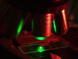
Technique in brief
Laser scanning confocal microscopy (LSCM)is a system used in epifluorescence and reflected light imaging. A finely focused beam of laser light is scanned across a sample and the resultant light emitted passed through a pinhole aperture to exclude any out of focus light. The image thus produced includes only the plane of focus for a given objective lens (hence a clearer image). Additionally, by varying the stage height, a series of sequential images through the thickness of a single sample can be collected and used to project a three-dimensional image.
A LSCM will typically include at least 3 different wavelength lasers which can be used simultaneously or sequentially. For imaging fluorescence in a sample this means multiple stains (fluorophores) can be used to highlight specific features of interest.
Current instrumentation
The current LSCM at the AAC is a BioRad Radiance 2000 with the following configuration:
Laser configuration
Argon ion - 488nm(14mW); 514nm(11mW)
Green HeNe - 543nm(1.5mW)
Red laser diode - 638nm(5mW)
Emission Filters
Detector (photomultiplier - PMT) Emission filters (wavelength in nm)
1 Open; 488/10; 500LP; 515/30; 530/60;
2 Open; 515/30; 530/60; 570LP; 590/70; 600/50; 600LP
3 Open; 660LP
It is based around the light optics of a Nikon E600 upright microscope with tungsten transmitted and reflected light sources and a mercury lamp. Current lenses available are; 10, 20 and 40x (cover slip corrected); 50 and 100x (air); 60x oil immersion and 40x (water immersion).
Applications
LSCM is used in many applications for imaging biological specimens. Although it cannot improve on magnifications available to conventional fluorescence microscopy the sharper, clearer images obtained greatly improving image quality. Also, as the system acquires images digitally, timed images may be automatically generated. In samples that are relatively transparent to the lasers the ability to create sequential images through the depth of a specimen (“z-series or stacks) produce 3-dimensional information provides real spatial context to the features of interest. Opaque materials can also be imaged this way, essentially recording reflected light. Thus it is also a useful technique in material sciences for modelling material in 3D.
Sample requirements
For fluorescence imaging specimens need by fixed and stained using fluorphores that will not only attach (label) to the relevant feature of interest but are excited by and emit at wavelengths appropriate to the configuration of lasers and detector filters available.
Samples for reflected laser light applications need be of a size suitable to be viewed on a conventional microscope.